Home
Uncategories
Shoulder Muscles Diagram Posterior / Muscles Moving Shoulder Posterior View Diagram Quizlet : Although anchored in the neck, their primary functions are to move the shoulder blades and support the arms.
Shoulder Muscles Diagram Posterior / Muscles Moving Shoulder Posterior View Diagram Quizlet : Although anchored in the neck, their primary functions are to move the shoulder blades and support the arms.
Shoulder Muscles Diagram Posterior / Muscles Moving Shoulder Posterior View Diagram Quizlet : Although anchored in the neck, their primary functions are to move the shoulder blades and support the arms.. Middle fibres are major abductors, anterior fibres flexion, posterior fibres extension Explore and learn the muscles of the shoulder with our 3d interactive anatomy muscle atlas. Deltoid tuberosity on humerus action: This diagram depicts shoulder muscle diagram. In the front of the neck, the.
The following is an overview of the shoulder muscle anatomy. Identifying muscles of the rotator cuff: Find out in this anatomy of the shoulder quiz. Learn vocabulary, terms, and more with flashcards, games, and other study tools. Joint pain exercises anatomy of neck and shoulder pain shoulder and clavicle anatomy posterior shoulder anatomy pain shoulder tendon pain 17 muscles of the shoulder shoulder skeleton anterior shoulder tendons anatomy shoulder bone structure shoulder pain bursitis shoulder.
Best Shoulder Exercises For You from www.pilates-back-joint-exercise.com These muscles give the sides of the neck their shape. A muscle contracts to move bones; Deltoid tuberosity on humerus action: Start studying posterior shoulder muscles. Although anchored in the neck, their primary functions are to move the shoulder blades and support the arms. Teres minor, supraspinatus, infraspinatus and subscapularis. Muscles of the upper arm and the shoulder blade. Learn vocabulary, terms, and more with flashcards, games, and other study tools.
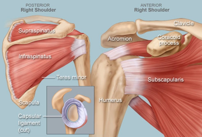
The shoulder anatomy includes the anterior deltoid, lateral deltoid, posterior deltoid, as well as the 4 rotator cuff muscles.
The tendons are the attachment of the muscle to the bone. Parts of the right shoulder blade: The shoulder anatomy includes the anterior deltoid, lateral deltoid, posterior deltoid, as well as the 4 rotator cuff muscles. Learn more about the other educational. In human anatomy the shoulder joint comprises the part of the body where the humerus attaches to the scapula the head sitting in the glenoid cavity. Deltoid tuberosity on humerus action: The shoulder is a complex combination of bones and joints where many muscles act to provide the widest range of motion of any part of the body. Anatomy • free medical books. Arm flexion (anterior), arm extension (posterior), and arm abduction (lateral) origin lateral 1/3 of clavicle, acromion, and spine of scapula. Teres minor, supraspinatus, infraspinatus and subscapularis. The shoulder joint also known as the glenohumeral joint is the main joint of the shoulder. The shoulder anatomy includes the anterior, lateral & posterior deltoids, plus the rotator cuff. Anatomy of the rotator cuff.
The following is an overview of the shoulder muscle anatomy. Start studying muscles of the posterior shoulder. These muscles form the outer shape of the shoulder and underarm. The muscles in the shoulder aid in a wide. Explore and learn the muscles of the shoulder with our 3d interactive anatomy muscle atlas.
Frozen Shoulder Adhesive Capsulitis Treatment Prevention from stretchcoach.com It causes pain in the area just outside the joint. These muscles form the outer shape of the shoulder and underarm. This diagram depicts shoulder muscle diagram. Learn their origins/insertions, functions & exercises. Muscles of anterior (flexor) compartment of arm, their origin, insertion, action/s and nerve supply are as follows superior ulnar collateral branch of brachial artery. Start studying muscles of the posterior shoulder. Shoulder joint anatomy chart chartex shoulder joint anatomy chart illustrates bones, ligaments, tendons and muscles from anterior, lateral, superior and posterior aspects. These muscles give the sides of the neck their shape.
All these pictures presented are printable shoulder muscle diagram resources.
Human anatomy diagram shoulder anatomy shoulder muscles shoulder muscles and chest. Muscles of anterior (flexor) compartment of arm, their origin, insertion, action/s and nerve supply are as follows superior ulnar collateral branch of brachial artery. Muscles of the upper arm and the shoulder blade. Arm flexion (anterior), arm extension (posterior), and arm abduction (lateral) origin lateral 1/3 of clavicle, acromion, and spine of scapula. These muscles give the sides of the neck their shape. Deltoid tuberosity on humerus action: Learn vocabulary, terms, and more with flashcards, games, and other study tools. These muscles form the outer shape of the shoulder and underarm. The following is an overview of the shoulder muscle anatomy. Shoulder girdle laterally (spine of scapula, acromion, some clavicle) inserts: Plus, exercises for training them. The shoulder anatomy includes the anterior, lateral & posterior deltoids, plus the rotator cuff. Learn more about the other educational.
Muscles of anterior (flexor) compartment of arm, their origin, insertion, action/s and nerve supply are as follows superior ulnar collateral branch of brachial artery. Find out in this anatomy of the shoulder quiz. Anatomy of the rotator cuff. The main shoulder muscles are trapezius, deltoid, pectoralis major and 4 rotator cuff muscles: On the anterior side of the shoulder, the coracobrachialis, serratus anterior, pectoralis major, and pectoralis minor muscles work as a group to flex and adduct the scapula and humerus anteriorly toward the sternum.
Shoulder Human Anatomy Image Function Parts And More from img.webmd.com It causes pain in the area just outside the joint. Middle fibres are major abductors, anterior fibres flexion, posterior fibres extension Ebraheim's educational animated video describes muscle anatomy of the shoulder girdle and anatomy of the shoulder joint.anatomy of the shoulder muscles a. The muscles of the upper arm are responsible for the flexion and extension of the forearm at the. Numerous muscles help stabilize the three joints of. Muscles of anterior (flexor) compartment of arm, their origin, insertion, action/s and nerve supply are as follows superior ulnar collateral branch of brachial artery. Formerly called tendinitis, this is inflammation or irritation of a tendon that attaches to a bone. Last update september 3, 2020.
In the front of the neck, the.
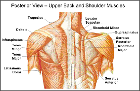
Arm flexion (anterior), arm extension (posterior), and arm abduction (lateral) origin lateral 1/3 of clavicle, acromion, and spine of scapula. In the front of the neck, the. These muscles give the sides of the neck their shape. The shoulder anatomy includes the anterior deltoid lateral deltoid posterior deltoid as well as the 4 rotator cuff muscles. Major muscles back muscles shoulder muscles supraspinatus muscle back workout routine sternocleidomastoid muscle muscle diagram body diagram latissimus dorsi. The main shoulder muscles are trapezius, deltoid, pectoralis major and 4 rotator cuff muscles: A muscle contracts to move bones; Find out in this anatomy of the shoulder quiz. Middle fibres are major abductors, anterior fibres flexion, posterior fibres extension The intrinsic muscles of the posterior group include the deltoid, teres major and the muscles of the rotator cuff. Starting point the muscles are the supraspinatus fossa on the surface, and mounting position. Ebraheim's educational animated video describes muscle anatomy of the shoulder girdle and anatomy of the shoulder joint.anatomy of the shoulder muscles a. Start studying muscles of the posterior shoulder.
The muscles of the shoulder are associated with movements at the shoulder joint shoulder muscles diagram. Human anatomy diagram shoulder anatomy shoulder muscles shoulder muscles and chest.

0 Comments:
Posting Komentar